As many of you know, and perhaps many of you don’t know, behind the scenes we have about 28 million specimens and several lab spaces where we investigate the diversity of life on our planet. The Project Lab is just a glimpse into some of the research that happens at the Academy. In the Project Lab, we have two stations where we can take high resolution images of tiny, microscopic ones (tiny seeds, insects, etc.) to large macroscopic specimens (fish, lizards, etc.). We take photos of specimens for many reasons, but most importantly is to communicate with others online or in publications.
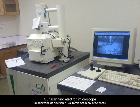
Lately, I’ve been working at an imaging station that you can’t see though the windows of the Project Lab. I’m talking about our scanning electron microscope (SEM). Our SEM is located on the bottom level of the building since it needs to be on stable ground to prevent movement of the microscope. The SEM is a type of microscope that uses electrons, instead of light, to create an image and is able to produce images at a very high magnification. These images tell us about the shape and texture or topography of different types of specimens. Some examples of specimens that biologists may image with an SEM include pollen grains, insect and spider genitalia, insect eyes, etc. As for the nudibranch lab at the Academy, we use the SEM to take images of a part of a nudibranch called the radula.
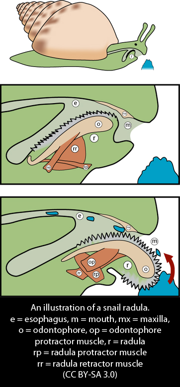
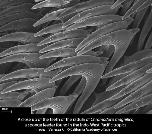
So what is a radula? The radula is the feeding structure of most mollusks (snails, slugs, squid, etc.). It is a sheet or ribbon-like structure that is made up of teeth that are made out of a substance called chiton. The radula is located inside the mouth-end of the mollusk and then comes out of the mouth to be used in scraping, cutting up or grasping food. In nudibranchs (and other mollusks), the radula is often used in species identification, so we often find ourselves cleaning and preparing nudibranch radular teeth under the microscope to take their picture. Although I did want to be a dentist when I was in the third grade, I never could have imagined that I'd be a slug dentist when I grew up!
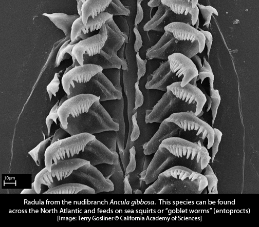
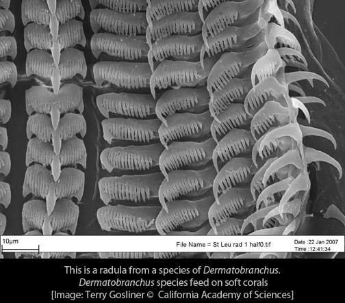
For many groups of nudibranchs, the radula will look different based on the species it came from. Major groups of nudibranchs will have similar looking radulae, and then within that group you will see variations. One reason for different types of radulae and different shapes of teeth is that different nudibranchs (and other mollusks) have different types of prey.
Let’s take a look at some nudibranch radulae images taken with an SEM...
This is just a small slice of the diversity of radulae found in the mollusk world. I'm going to get back to documenting some of it, so that's all for now! Till next time...
Vanessa Knutson
Project Lab Coordinator and Graduate Student